
Impedance audiometry, middle ear analysis, acoustic reflex testing
Tympanometry and Impedance Audiometry
A rapid objective measure of how the eardrum and middle ear move, detecting fluid, negative pressure, perforation and ossicular problems within seconds per ear.
What this assessment is
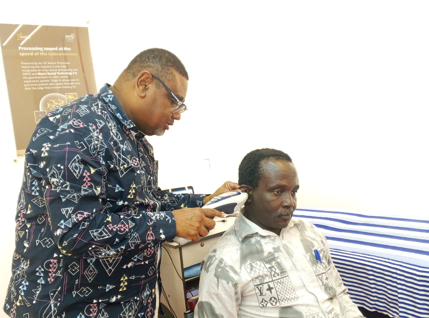
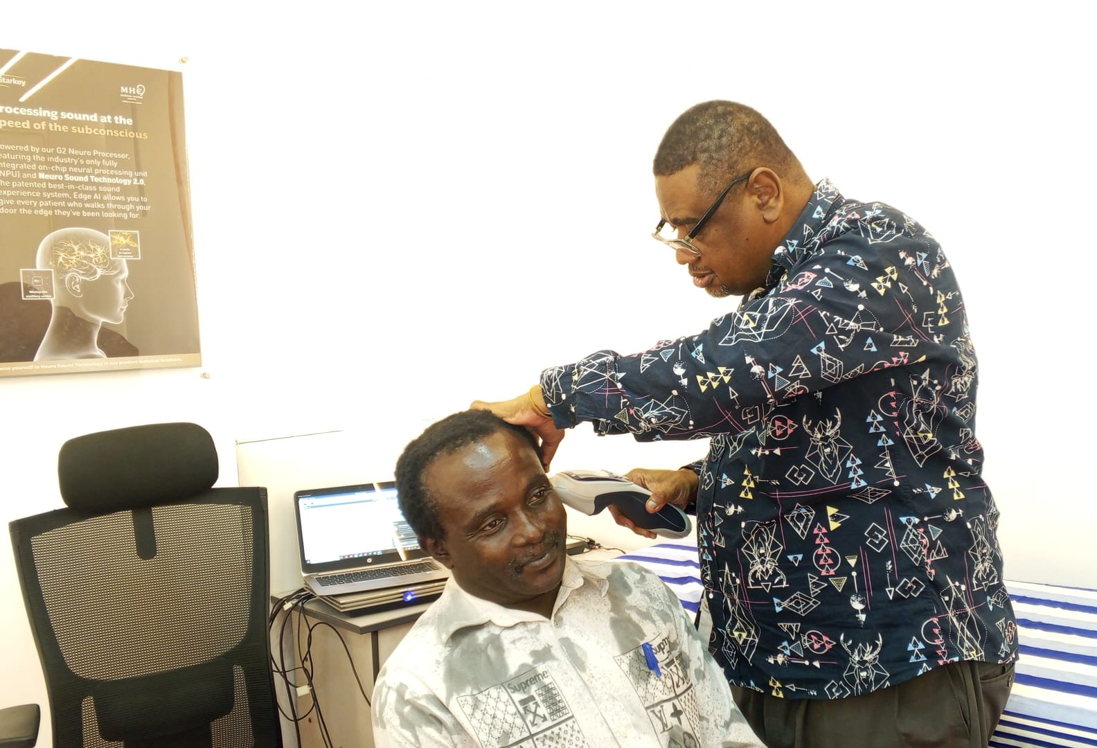
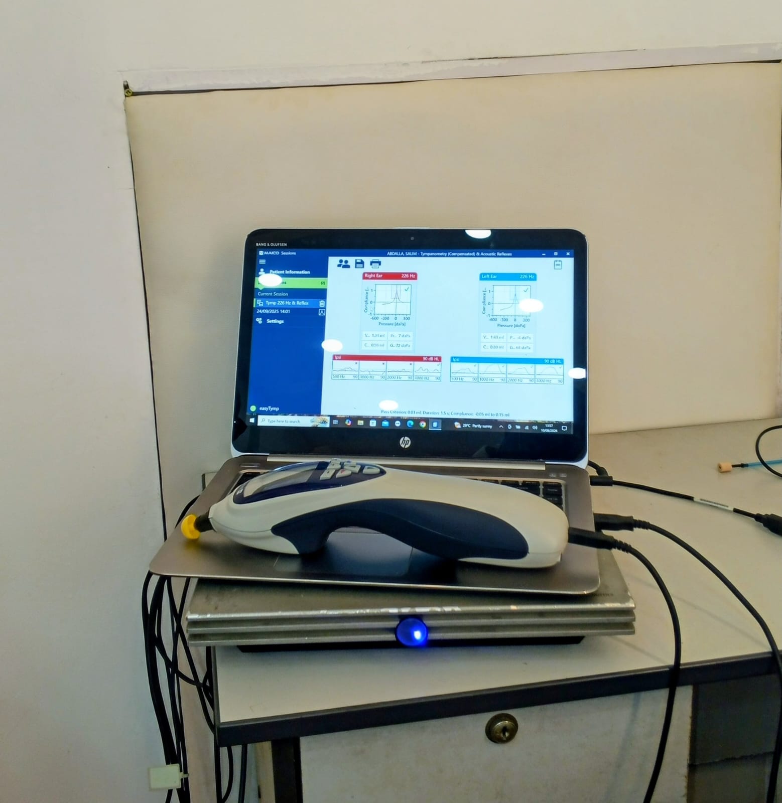
Where pure tone audiometry describes what you hear, tympanometry explains part of the reason. A soft probe tip seals the ear canal and varies the air pressure while a 226 Hz tone is delivered. The instrument measures how much sound energy the eardrum admits at each pressure and draws a tympanogram. A healthy middle ear peaks near zero decapascals; fluid, blocked eustachian tubes, perforations and disrupted ossicles each produce their own recognisable trace. At Mombasa Hearing Centre this test is run on both ears in the same sitting, and the results are captured directly onto the clinical software for the file.
Why it matters
- It identifies middle ear fluid before it causes obvious symptoms, which is critical in children.
- It confirms or rules out a conductive component objectively, supporting the air and bone results from audiometry.
- Acoustic reflex thresholds add information about the middle ear muscles, the auditory nerve and the brainstem pathway.
- It requires no response from the patient, so it can be performed on infants, elderly patients with dementia, and anyone unable to cooperate.
At the centre
How it looks in practice
Photographs taken at Mombasa Hearing Centre during real clinical sessions.
Step by step
How the assessment is performed
Each stage follows established audiological protocol, and you are told what is happening before it happens.
- 1
Otoscopy first
The canal is inspected to confirm there is no wax occlusion or discharge and that a safe seal can be achieved.
- 2
Probe placement and seal
A soft disposable tip sized to your ear is placed at the canal entrance. You will be asked to stay still and avoid swallowing, talking or yawning for a few seconds, since any of these change middle ear pressure.
- 3
Pressure sweep and tympanogram
Pressure is swept from positive to negative, typically +200 to -400 daPa, while admittance is measured. The instrument reports peak pressure, static compliance and ear canal volume.
- 4
Acoustic reflex thresholds
Louder tones at 500, 1000, 2000 and 4000 Hz are presented to trigger the stapedius muscle contraction. Present reflexes at normal levels are reassuring; absent or elevated reflexes direct further investigation.
- 5
Interpretation and reporting
Traces are classified and read alongside the audiogram. The audiologist explains what the shape of your tympanogram means and whether an ENT opinion is needed.
How to prepare
- Let us know if you have a perforated eardrum, a grommet, or recent ear surgery, as the protocol is adjusted.
- Avoid using ear drops on the morning of the test unless prescribed.
- Stay still and quiet during the few seconds of recording for a clean trace.
Understanding your results
- Type A, normal
- A clear peak near zero daPa with normal compliance. The middle ear is ventilated and mobile.
- Type B, flat
- No peak. With a normal canal volume this usually indicates fluid behind the eardrum; with a large volume it suggests a perforation or a patent grommet.
- Type C, negative pressure
- The peak is shifted negative, pointing to eustachian tube dysfunction, often after a cold, allergy or air travel.
- Shallow or very deep peaks
- Reduced compliance can indicate a stiff or fixed ossicular chain, while an unusually high peak can indicate a disarticulation or a very flaccid eardrum. Both warrant an ENT referral.
FAQ
Common questions
Is any liquid or instrument put into my ear?
No. Only a soft probe tip at the canal entrance, and only air pressure and sound are used.
Can it be done if my eardrum is perforated?
Yes, and it is useful. The measured canal volume helps confirm the perforation and estimate its effect.
Does it replace the audiogram?
No. Tympanometry tests the middle ear mechanism, not your hearing sensitivity. The two tests answer different questions and are read together.
Explore
Other tests we perform
Pure Tone Audiometry
The reference test for hearing. It maps the quietest sounds you can detect across the speech frequencies and shows whether a loss sits in the outer, middle or inner ear.
Paediatric Hearing Assessment
Age appropriate testing for babies and children, using play, toys and visual rewards to obtain reliable thresholds long before a child can describe what they hear.
Brainstem Evoked Response Audiometry
An objective test of the hearing nerve and brainstem pathway. Small surface electrodes record the brain's automatic response to sound while the patient rests or sleeps quietly.